
One of the ways technology has empowered people is by providing easy access to knowledge. The internet has become a veritable trove of information we can explore with just a few taps or clicks. However, not all internet sources are reliable. Many have not been checked for accuracy, while others are written with bias or contain misleading content. While some of these are easy to countercheck, there are some that could lead to serious consequences. In particular, inaccurate medical information can compromise one’s health and even prove fatal in worst-case scenarios. Yet, there are many people who rely on the Internet for health-related concerns.
How Accurate Is Medical Information Online
While there are independent sites that provide reliable medical information, some studies show that medical information from the internet can often be inaccurate. A study on the accuracy of online information on infant sleep safety shows that only 43.5 percent of the 1,300 websites analyzed provided accurate content based on the recommendations of the American Academy of Pediatrics (AAP). Another study conducted to determine the educational quality of YouTube videos for asthma shows that most of them were a poor source of accurate health care information.
Still, the free access to these sources of information attracts people who are looking for more convenient and affordable ways to understand their condition. After all, the internet is free, but medical consultations often come with hefty fees.
It’s virtually impossible for the medical community to stop people from accessing sources on the web. That said, some actions can be taken to ensure the accuracy of medical content they access. Doctors and health practitioners can lead their patients to trustworthy sites that ensure the veracity and accuracy of all the content they publish. Medical illustrators also play a key role in conveying accuracy in information.
Enhancing the Accuracy of Medical Content Through More Precise Illustrations
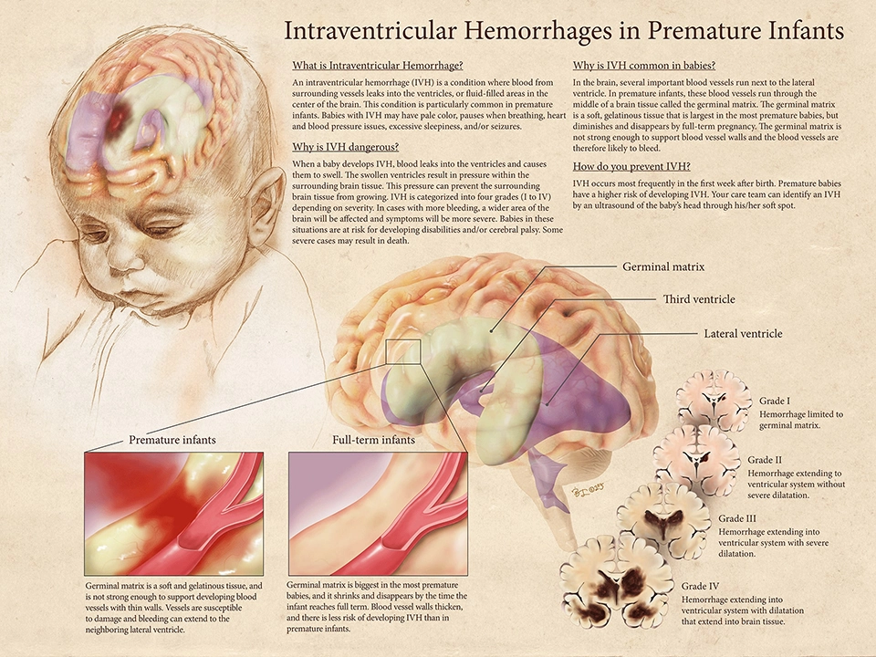
Intraventricular Hemorrhages in Premature Infants
As medical illustrators, we constantly seek to augment the limitations of researchers and publishers in presenting their studies. We create clearly defined and highly detailed illustrations to enrich medical content and make them more engaging to the target audience. One of our recent works, the Intraventricular Hemorrhages in Premature Infants demonstrates how we addressed a need for more accurate medical illustration.
Dr. Aarti Raghavan, a neonatologist at the University of Illinois Hospital, expressed how the lack of an adequate medical illustration prevented her from showing images to her colleagues and the patients’ family members to explain intraventricular hemorrhage (IVH). This condition is characterized by blood leaking from the surrounding vessels into the lateral ventricles inside the brain. Because the blood vessels in the brain are not yet fully developed, premature infants born before 28 weeks of gestation have higher risks of IVH.
Most illustrations of IVH on Google feature the lateral ventricles of adults. However, since the lateral ventricle is not fully developed to resemble that of adults until the 31 week of gestation, the images found online do not represent that of neonates.
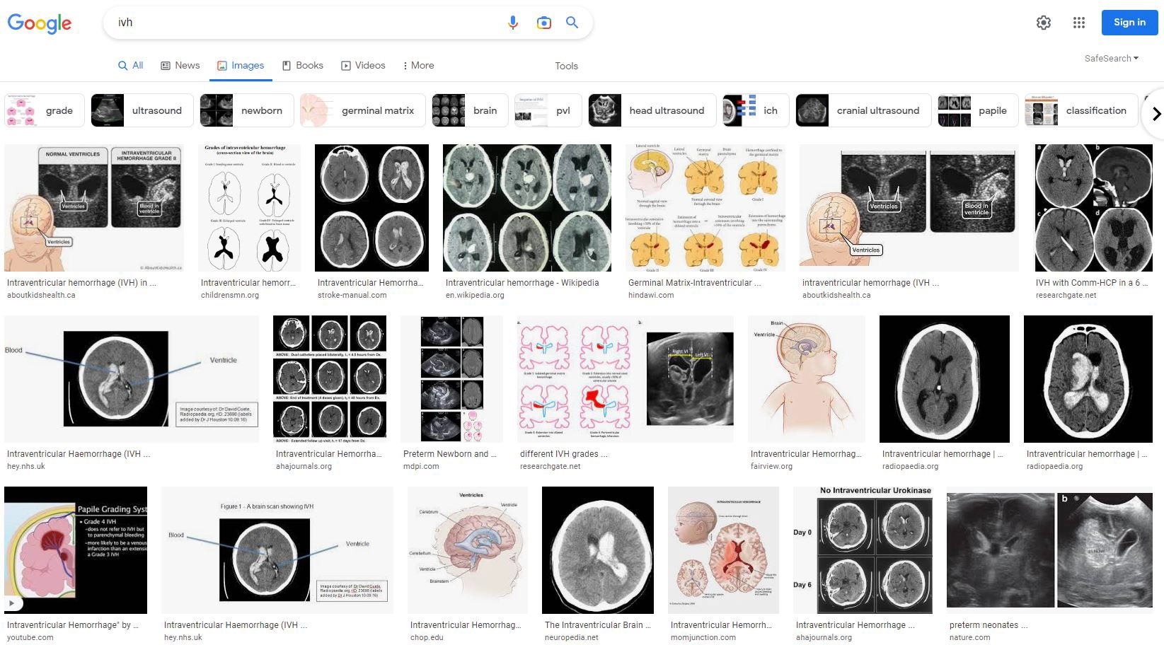
Screen capture of a Google image search on “IVH”
Finding Solutions to Imaging Limitations
The lack of images that accurately represents the brains of neonates derives mainly due to difficulty in imaging their brains. Since their brain is still developing, their radiosensitivity and lifetime radiation risks are much higher than adults. Thus, exposing them to imaging tools such as CT scans puts them at risk of severe conditions such as cancer. An MRI offers a safer and non-invasive imaging tool, but its sensitivity to motion is a challenge for pediatric scans.
To create an accurate illustration of infant brains, we conducted our own research and referenced a study of a volumetric imaging of a 3D MR of fresh fetal brain specimens from legally aborted babies with gestational age (GA) of 7 to 28 weeks. The image showed both the lateral ventricle and the germinal matrix, which were crucial for constructing the illustration..
We used the frontal, superior, lateral, and inferior view of the imaging results as a guide to reconstruct the brain of a 28-week old infant on ZBrush, a 3D sculpting software. To create a 3D static image, we aligned the position of the lateral ventricle with the infant brain three-dimensionally using 3D software, Autodesk 3D Studio Max. Using a real reference of a baby, we aligned the brain with the sketch-style illustration of the baby.
Now, we are proud to share the only IVH image that accurately represents the brain of an infant with a 28-week gestational age. Aside from giving audiences a more accurate illustration of the brain map of those suffering from the highest risk of IVH, this illustration also helps parents understand their babies’ condition. This is an attempt to avoid overwhelming the parents who are already under tremendous anxiety and stress from their newborn’s health condition.
Working Hand in Hand to Boost Accuracy of Medical Information Online
The internet will likely continue to be the primary source of information for the public. While it poses challenges, the medical community can work hand in hand to uplift the quality of knowledge that people gain from online sources.
Healthcare professionals can partner with medical illustrators who can create accurate content that supplements medical information. They can communicate the problems they seek to address and collaborate with illustrators on the most appropriate solutions. Being well versed in the fields of science and medicine, medical illustrators are more than capable of translating ideas, facts, and data into highly detailed images and engaging infographics.
If you need high-science graphic content, visit maomiyamoto.com to discuss your needs and get a quote.