Project Details
- Client :
University of Illinois at Chicago - What We Did :
Infographic Creatiion - Tools Used :
Photoshop, Illustrator - Completed on :
December 2015 - Skills :
Digital painting - Audience :
Patients and their parents
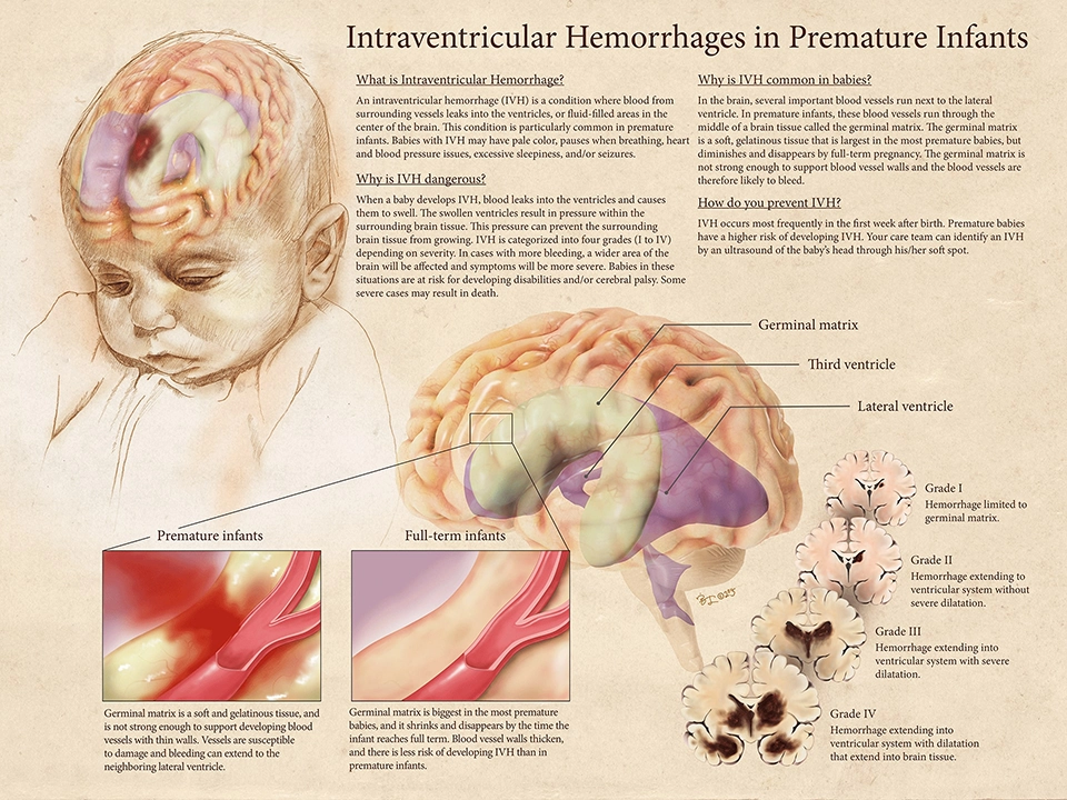
Intraventricular hemorrhages in premature infants
Intraventricular hemorrhage (IVH) is extremely common in premature infants, and is caused by the unique undeveloped structure of the neonatal brain. Swiftly educating the patient’s parents is important because prompt response is crucial to prevent the condition to cause sever dysfunction in the future. Only ultrasound brain images or inacurate illustration of an infant brain were available for the pediatritians to present to the patients, which was either too hard or incorrect for education. Lack of accurate illustration is due to higher risk and difficulty is involved in exposing the neonates to brain scan, leaving no reference for content development. Our client at the NICU department at the University of Illinois Hospital tasked us to create an educational piece that can be used to achieve better communication around IVH.
Utilizing our research skills, we reconstructed the 3D brain structure of neonates utilizing an academic research that ethically conducted a brain scan of a deceased neonate. This became the one and only accurate representation of a neonatal brain, helping pediatritians across the globe to educate parents from different background and education level in understanding the mechanism of their child’s condition and making a and better life for thier child.
Step 1: Science download
During our deep dive, we discovered the problem to be two folds: 1. many resources are MRI data, making it harder for the audience to understand. 2. medical illustration represented adult ventricles, while infant brain is visually different from the adult's, making the information inaccurate.
Step 2: Sketches
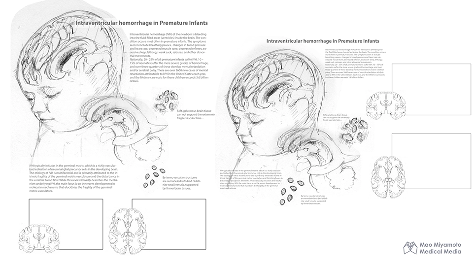
While conducting the research, we compiled necessary information for the patient to make an informed decision and created various compositions, making sure that the information flows intuitively. Unlike many illustration we found online, we centered a story around germinal matrics, a leaky gelatinous structure only present in premature infant's brain. We also aimed to show the structure from multiple angles to provide better spacial orientation to the patients.
Step 3: Building Assets
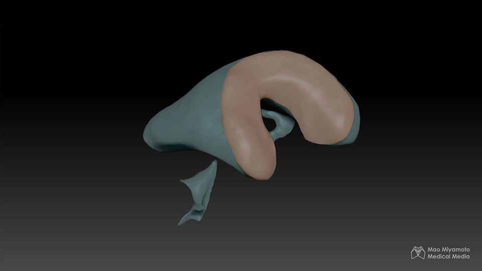
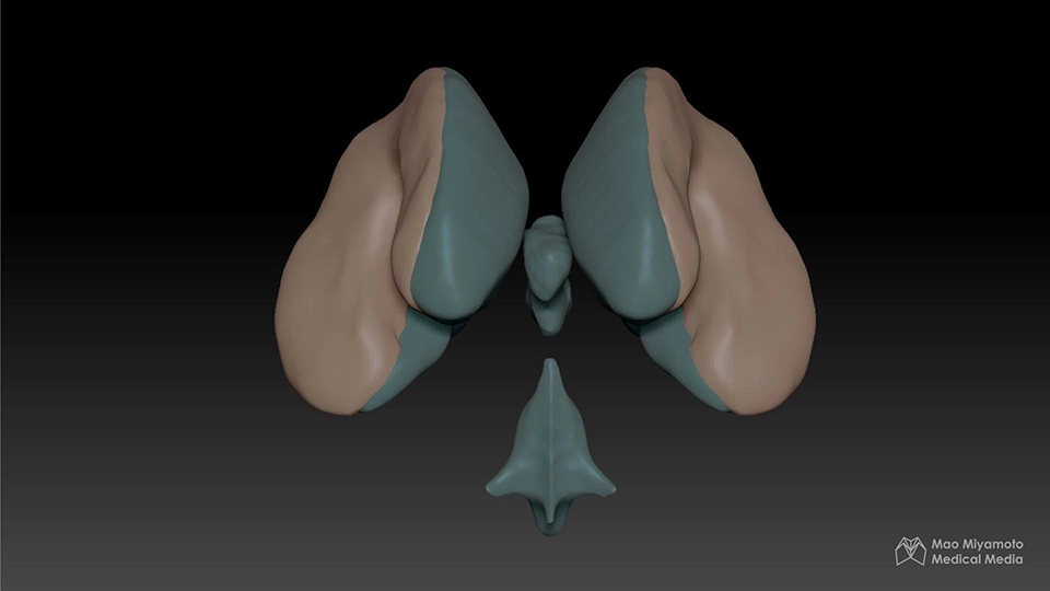
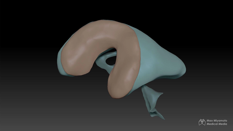
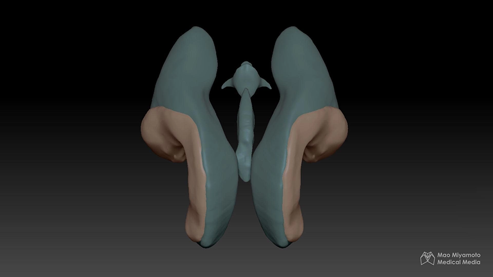
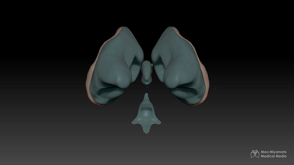
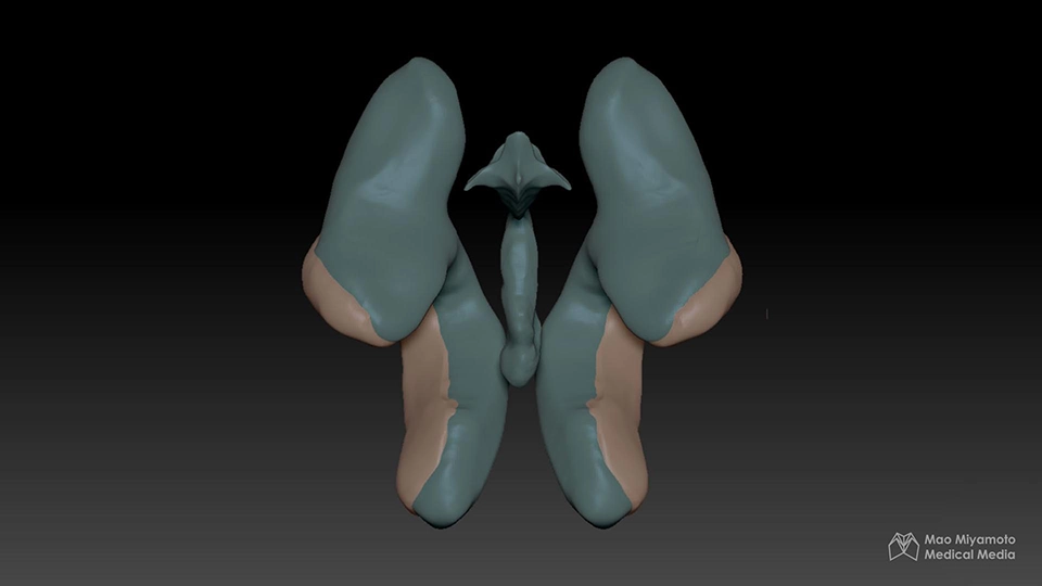
Conducting an accurate scanning of a baby’s brain, through X-ray or CT come with severe radiation and cancer risks, while MRI can also be hard as it is sensitive to motion. Using the academic article Volumetric Analysis of the Germinal Matrix and Lateral Ventricles Performed Using MR Images of Postmortem Fetuses, we reconstructed an accurate 3D representation of the neonate’s ventricles with germinal matrics, the first and only 3D resource you can find.
Final Deliverable